 By Dr Jean-Pierre Lin, Evelina London Children’s Hospital, London, UK
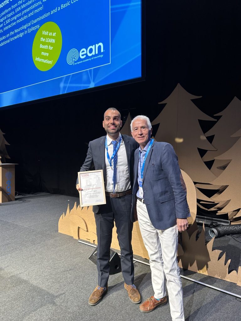
The award of the Dystonia Europe David Marsden Prize to Dr Stavros Tsagkaris represents a significant recognition of the importance of our work in investigating brain activity patterns in childhood and adolescent dystonias1, 2 representing a 20-year project to carefully unravel the mechanisms of dystonia in children and young people.
Setting up a children’s dystonia diagnostic and management service:
In 2005 the embryonic Complex Motor Disorders Service (CMDS) at the Evelina London Children’s Hospital set up a Multidisciplinary Team (MDT) to deliver a Deep Brain Stimulation (DBS) Service for children with dystonia and other movement disorders in collaboration with neurosurgical colleagues: Mr Richard Selway, Prof. Keyoumars Ashkan and latterly Mr Harutomo Hasegawa from the King’s College Hospital Neurosurgery Department.
In 2008 Dr Jean-Pierre Lin was awarded a Guy’s and St Thomas’ Charity New Services and Innovation Grant G060708 for pump-priming support to build a dedicated CMDS MDT Team, leading to the current clinical-research structure.
Dystonia Brain Function:
Given the relative rarity of individual causes of dystonia in childhood the CMDS MDT set out to gather the broadest possible information that could result in a better understanding of the impact of dystonia on function, comfort, growth and development and well-being in children and young people, by collecting very detailed clinical information on each child. Together with dedicated scales to measure function, dysfunction and dystonia alongside genetic, metabolic, imaging and neurophysiological parameters.
Neurophysiology of dystonia:
Dr Verity McClelland spearheaded neurophysiological testing of the central motor conduction time (CMCT)4with transcranial magnetic stimulation (TMS) and evaluation of the sensory pathways from somatosensory evoked potentials (SEP)4. This was followed by simultaneous brain and muscle activity patterns during simple manual tasks5.
Brain Activity Patterns in childhood dystonia:
Two imaging techniques were used to study the brain of children with dystonia: MRI brain imaging, led by Dr Daniel Lumsden, produces detailed structural brain images. Fluorodeoxy- Glucose Positron Emission Tomography brain scans fused with CT imaging (FDG-PET-CT), led by Dr Jean-Pierre Lin and Prof. Alexander Hammers, allow us to study brain activation patterns in dystonia.6,7
How investigations and clinical evaluation are combined in assessing childhood dystonia:
Dr Stavros Tsagkaris studied the similarities and differences in regional brain glucose activity (i.e. glucose uptake and metabolism) in quietly awake children with dystonia which has shone a new light on the similarities and differences between different causes of dystonia when compared to healthy controls without dystonia.
This international multidisciplinary collaboration has enabled an understanding of direct brain activity assessment in childhood dystonias.
Future directions:
The very large comprehensive dataset collected by the CMDS team over the last 20 years will allow careful unravelling of the many factors underpinning childhood-onset dystonias by describing and following up brain activity patterns (glucose consumption). With this data we can track the impact of age, timing and aetiology (i.e. causes) of dystonia.
Future applications of FDG-PET-CT brain imaging will include ‘before’ and ‘after’ dystonia intervention imaging studies to evaluate the impact of advanced therapies such as deep brain stimulation or gene therapies on childhood brain activity patterns.
We intend to build on previous data7 to characterise individual brain energy consumption patterns to fully explore the impact of the many variables, singly and in combination, underlying the similarities and differences in dystonia, expressed in all age groups, and relative responsiveness to DBS and other advanced therapies.
Our aim is to generate new hypotheses regarding causal mechanisms of dystonia leading to new management strategies.
Acknowledgements:
Dr Jean-Pierre Lin supervised the clinical data collection; Professor Alexander Hammers, head of the King’s College London PET Centre at St Thomas’s Hospital, supervised the FDG-PET-CT analyses and Dr Eric Guedj of the Unveristé of Aix Marseille, France, kindly provided the healthy adult control FDG-PET-CT brain scan data.
Funding and support:
J-P Lin: Guy’s and St Thomas’ Charity New Services and Innovation Grant G060708; Dystonia Society UK Grants 01/2011 and 07/2013 and Action Medical Research GN2097; GSTT Charity Complex Motor Disorders Service Research and Education Fund A13.
A Hammers: Wellcome EPSRC Centre for Medical Engineering at King’s College London (WT 203148/Z/16/Z) and also from the Department of Health via the National Institute for Health Research (NIHR) comprehensive Biomedical Research Centre award to Guy’s & St Thomas’ NHS Foundation Trust in partnership with King’s College London and King’s College Hospital NHS Foundation Trust.
- McClelland: is currently supported by a Clinician Scientist Fellowship from the Medical Research Council UK (MRP0068681) and by a Continuation Funding grant from the Rosetrees Trust (CF-2021-2-112) and was previously supported by a postdoctoral clinical research training fellowship from the Medical Research Council UK (MRP0068681) and a project grant from the Rosetrees Trust (A1598).
- Stavros Tsagkaris, Eric K C Yau, Verity McClelland, Apostolos Papandreou, Ata Siddiqui, Daniel E Lumsden, Margaret Kaminska, Eric Guedj, Alexander Hammers, Jean-Pierre Lin.
Metabolic patterns in brain 18F-fluorodeoxyglucose PET relate to aetiology in paediatric dystonia. Brain, Volume 146, Issue 6, June 2023, Pages 2512–2523, https://doi.org/10.1093/brain/awac439
- TA Szyszko, JT Dunn, MJ O’Doherty, L Reed, JP Lin.
Nuclear medicine communications. 2015, 36 (5), 469-476
- VM McClelland, A Valentin, HG Rey, DE Lumsden, MC Elze, R Selway, G Alarcon, JP Lin.
Journal of Neurology, Neurosurgery & Psychiatry 2016, 87 (9), 958-967
- Verity M McClelland, Doreen Fialho, Denise Flexney-Briscoe, Graham E Holder, Markus C Elze, Hortensia Gimeno, Ata Siddiqui, Kerry Mills, Richard Selway, Jean-Pierre Lin.
Clinical Neurophysiology 2018, 129 (2), 473-486
- VM McClelland, Z Cvetkovic, JP Lin, KR Mills, P Brown
Abnormal patterns of corticomuscular and intermuscular coherence in childhood dystonia
Clinical Neurophysiology 2020, 131 (4), 967-977
- H Gimeno, JP Lin
The International Classification of Functioning (ICF) to evaluate deep brain stimulation neuromodulation in childhood dystonia-hyperkinesia informs future clinical & research …priorities in a multidisciplinary model of care.
European Journal of Paediatric Neurology 2017, 21 (1), 147-167
- SA Shah, P Brown, H Gimeno, JP Lin, VM McClelland
Frontiers in neurology 2020, 11, 825
