For patients living with dystonia, deep brain stimulation (DBS) is an established, safe and effective treatment option.1
DBS is a surgical procedure during which two thin electrodes are inserted into the brain and conneted to a stimulator via an extension in the chest or abdomen.2 The treatment works by delivering targeted electrical pulses to the brain, blocking the signals that cause involuntary muscle contrations in patients with dystonia and easing symptoms, such as tremor and pain.2
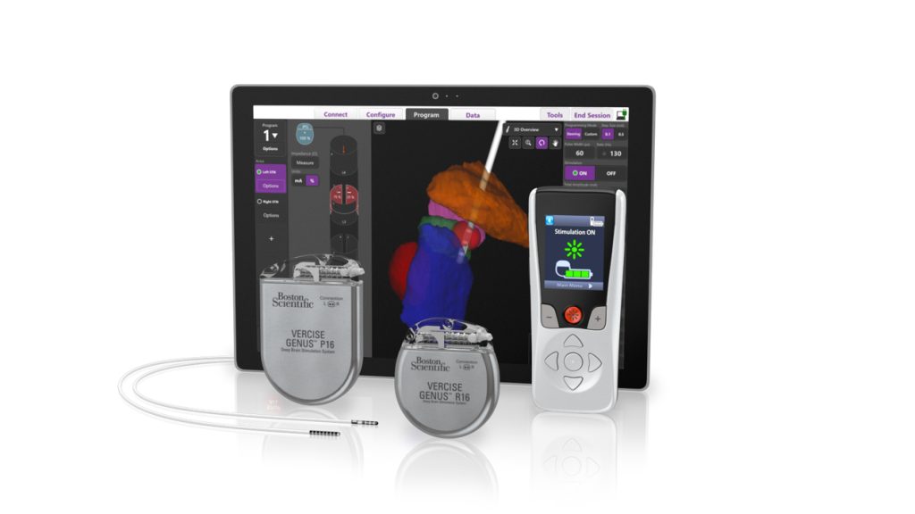
Over the 35 years since DBS was first developed, 3 considerable improvements have been made to enhance the technology. In the early days, clinicians did not have a lot of information to work with during the programming – relying on a ‘trial and error’ basis when placing the electrodes. In more recent years, as the progress of this treatment has continually improved, one ground-breaking development has been the introduction of visualisation software, such as the new Stimview-XT from Boston Scientific, which allows neurosurgeons to ‘see’ electrode placement in relation to a patient’s specific brain anatomy.
Armed with the knowledge of how certain lead placement and contact decisions will impact the patient’s symptoms, it is now possible to achieve a level of precision never experienced before, greatly factoring into the ongoing improvement of personalised treatments for each individual patient. Professor Volker A Coenen, University Medical Center Freiburg, who has been instrumental in the development of advanced imaging for more than 10 years, explains the significance of the software: “Visualisation software helps to join the dots in DBS. It is being utilised during the implant surgery to optimise placement and programming and help deliver the best outcomes for the patients.”
![]()
Seeing the communication gap with an image
With the visualisation tool available, diverse clinical teams – who don’t often have the chance to collaborate – can achieve better planning, programming and patient care because they are aligned – literally seeing the same results: something medical teams have long been striving for. “We’ve been wanting to go in this direction for over 20 years. It is a dream come true,” explains Prof Coenen. Creating the 3D reconstruction of the patient’s brain anatomy is a complex process, which is made simple and clear on a screen for every team member to access, enabling them to fine- tune and personalise DBS for dystonia patients.
Not only can this software potentially improve the accuracy and outcomes of DBS, it is also enabling a more streamlined workflow between these clinical teams. Prof Coenen adds: “Visualisation is starting to be much easier. The neurologist can come to me with a USB stick and then I can do the simulation.” As the software becomes more straightforward to use, it is hoped that even more patients may benefit.
While visualisation software is primarily used to place electrodes correctly, it can also be used for post-operative review of DBS, where teams can again use imagery to speak the same language as they explore potential improvements. Prof Coenen discusses: “We always look at the post-operative position of our electrodes, in every case. We have cases where we could improve the situation for the patient by utilising the possibilities that visualisation software presents.”
Seeing is believing
While one of the main benefits of the software is that it allows teams, such as Prof Coenen’s, to offer a more personalised and accurate therapy, some patients can still feel daunted by the prospect of DBS. In Prof Coenen’s experience however, the benefits significantly outweigh any initial doubts or concerns: “The feeling of being scared wears off when they realise the benefits to them. It is a question of accommodating to the situation, then there is no reservation.”
For patients to best understand the process, “seeing is believing” says Prof Coenen, adding: “I can show the patient ‘this is your head and these are the electrodes and this is how I can move them around.’ They understand. Then I programme it and if it’s better they say: ‘oh wow, that’s what the electrodes do.’ We’ve always worked this way because patients appreciate being shown things on screen… some patients ask to see a photo of what is being done and this helps make it more real for them. They can comprehend what is happening,” adds Prof Coenen.
DBS is not an experiment
Summarising his discussion on DBS and visualisation software, Prof Coenen believes that patients should be as informed as possible, so that they can benefit from DBS conducted using visualisation software. His advice to patients: “Don’t feel that DBS is an experiment. It is going to help you; most patients are really helped by DBS. Go to an expert, have a consultation and ask questions so that you can understand if this might be the therapy you need. In dystonia, I have seen so many wonderful things happen for patients.”
Visualisation software is a long-awaited game changer for neurosurgeons performing DBS, helping them to achieve a level of precision that was once unattainable. It has created a common language amongst clinical teams, ensuring everyone is striving for the same result. As a patient with dystonia, you can feel confident that your clinical team is using first-class technology to fine-tune and personalise your DBS, with the ultimate aim of improving your quality of life. The software is designed to ensure that everyone is on the same journey, understanding the process together and treating with precision.

Prof Volker A Coenen
References
- Ortiz, R.M., Scheperjans, F. & Pekkonen, E. Deep brain stimulation for dystonia in Finland during 2007–2016. BMC Neurol. 2019;19:137.
- Dystonia UK. Deep brain stimulation. 2019. Accessed online on 24 March 2022. Accessed at: https://www.dystonia.org.uk/deep-brain-stimulation
- Gardner J. A history of deep brain stimulation: Technological innovation and the role of clinical assessment tools. Soc Stud Sci. 2013;43(5):707-728.
IMPORTANT INFORMATION: These materials are intended to describe common clinical considerations and procedural steps for the use of referenced technologies but may not be appropriate for every patient or case. Decisions surrounding patient care depend on the physician’s professional judgment in consideration of all available information for the individual case. Boston Scientific (BSC) does not promote or encourage the use of its devices outside their approved labeling. Case studies are not necessarily representative of clinical outcomes in all cases as individual results may vary.
CAUTION: The law restricts these devices to sale by or on the order of a physician. Indications, contraindications, warnings, and instructions for use can be found in the product labelling supplied with each device or at www.IFU-BSCI.com.
Products shown for INFORMATION purposes only and may not be approved or for sale in certain countries.
NM-1284901-AA © Boston Scientific International